Coagulated or clotted blood that resembles a gel-like structure is called a blood clot or a thrombus. The most common occurrence of a blood clot is at the site of a bleeding wound. Blood clots (thrombi) are healthy and life-saving when they perform the task of stopping a wound from bleeding. They can also be a cause of a life-threatening condition when formed in the blood vessels.
Clotting is the body’s natural response to blood vessel damage, which prevents blood from leaking out and protects the underlying tissue. Blood clots that block arteries and prevent the flow of oxygenated blood to an organ causing tissue damage (infarcts); this can, however, be dangerous. Breaking away of blood clots from the area they are meant to protect (called embolism) can lead to life-threatening conditions.
Understanding clot types for diagnosis
Blood clots formed in the arteries are known as arterial clots, and those formed in the veins are known as venous clots. Arterial clots show immediate signs and require emergency treatment, whereas, venous clots build up slowly, but still lead to serious complications. Deep vein thrombosis (DVT), however, occurs when clots are formed in a major vein deep inside the body.
When symptoms of blood clots do appear, some of them are the same as other diseases. A consultation with the doctor is essential to confirm blood clots as the cause of symptoms.
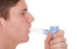
Diagnosis of blood clots
Diagnosis of blood clots is a tricky matter. As per the Centers for Disease Control and Prevention, almost 50% of those affected by DVT show no symptoms. It is best to call local emergency services if you experience a sudden shortness of breath; pressure in your chest; or difficulty in speaking, seeing, or breathing. The doctor or healthcare provider will evaluate the symptoms and run one of the following diagnostic tests to confirm the cause of the symptoms.
- Venous ultrasound: This is usually the first test conducted to confirm a venous blood clot. A Doppler ultrasound is used to visualize blood flow through the veins; however, in some cases, a venography or MR angiography is used to further confirm the findings of the ultrasound.
- D-timer test: This test measures the presence of a substance in the blood that is causing abnormal clotting.
- Cardiac biomarker test: This test evaluates the damage to the heart muscle and diagnoses a heart attack.
- Compression ultrasound: This is a non-invasive test that is useful in diagnosing DVT.
- Head CT scan: This is the primary test conducted to diagnose a stroke caused by a blood clot in the brain or skull.
- Chest CT Scan: This is conducted if the doctor suspects pulmonary embolism.
- Abdominal/pelvic CT scan: This diagnostic test is performed when a blood clot is suspected in the abdomen or pelvic region.
- MRI scan: This scan helps detect clots in blood vessels.
- Venography or angiography: These tests are catheterization methods that use a dye to detect clots.
- Echocardiography: This test captures images of the heart to detect embolism.
Treatment of blood clots
Arterial clots are treated through catheter-directed thrombolysis. This procedure essentially delivers clot bursting medication to the site of the clot. Blood-thinning medication is the normal treatment used for venous clots. For people with a high risk of developing venous clots, a vena cava filter is opted for.
As with any disease, prevention is better than cure. Here are a few self-care measures that mitigate the risk of developing blood clots:
- Avoid sitting for a long time
- Consume plenty of liquids while traveling
- Start exercising regularly, quit smoking, and maintain blood pressure levels